

The Detection and Impact of Surgical Smoke in the Operating Room
The operating theatre is a highly specialised environment focused on precision, sterility and the safety of patients and staff. However, the routine use of equipment such as tissue ablation tools, electrocautery tools and ultrasonic surgical devices consistently introduces an overlooked hazard to all people within the operating theatre. Surgical smoke is not simply just a visual hindrance or nuisance, but instead is a complex chemical biohazard generated by the thermal destruction of tissue. Even in many modern, highly regulated facilities, surgeons, nurses and patients are regularly exposed to surgical smoke, with it lingering directly within the perioperative zone (Malik et al., 2022).
Surgical smoke functions as an extreme chemical, biological and respiratory stressor, in which inhalation of the smoke forces the body to absorb toxic volatile organic compounds (VOCs) such as hydrogen cyanide, toluene, benzene, and carbon monoxide (Fencl, 2017) and other compounds like non-viable cellular material, viruses, and bacteria (Malik et al., 2022). Ongoing exposure to these VOCs not only triggers acute symptoms like headaches and airway irritation but also presents long-term risks of chronic pulmonary disease and cancer, with one study identifying nine main carcinogenic chemical compounds in surgical smoke (Liu et al., 2019). Understanding surgical smoke and when it is released in dangerous quantities is crucial for recognising why modern detection techniques, protective gear, and ventilation systems are so important in safeguarding the health of all those in the operating room.
This white paper explores the biological hazards of surgical smoke and the need for modern detection techniques in the operating room.
Chemical and mutagenic hazards of surgical smoke
Surgical smoke generated by several thermal tissue-destruction processes can significantly degrade the air quality in the operating theatre, creating a hazardous environment for the medical team. Clean air is fundamental to maintaining a safe surgical workplace, yet smoke generated during routine procedures and operations introduces a highly concentrated mixture of gases and particulates.
Research indicates that when tissue is thermally ablated, VOCs and known carcinogens are released, in which hazardous particles that are greater than 5 μm can deposit on the walls of the nose, pharynx, trachea and bronchus, whereas particles smaller than 2 μm are deposited in the bronchioles and alveoli (Malik et al., 2022), see Figure 1. Given that 77% of particles in surgical smoke are in the inspirable range, this is increasingly concerning to surgical staff and patients, as it poses a risk of alveolar congestion, interstitial pneumonia, bronchiolitis, emphysematous changes, and chronic inflammation (Hill et al., 2012). In many operating theatres, where there are continuous surgeries taking place with little time for ventilating the theatre, the levels of surgical smoke produced daily can be equivalent to 27-30 cigarettes (Hill et al., 2012). This is particularly a problem for nurses who spend more time in the theatre and are therefore “continuously exposed to hazardous surgical smoke” (Moon et al., 2021), an opinion evidenced by Ilce et al.’s (2017) research in which half of the nurses and doctors in their study acknowledged adverse symptoms from surgical smoke.
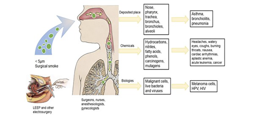
Figure 1.
Establishing a direct link between immediate patient wound contamination and surgical smoke requires further research; however, the presence of bioaerosols in surgical smoke poses serious safety risks to both the surgical team and patients. Under unmitigated smoky conditions, smoke can act as a live vector, carrying pathogenic cellular material; for instance, studies have shown that smoke produced from ablated breast tissue exhibits mutagenicity in a TA98 strain of Salmonella (Malik et al., 2022). Alongside this, another study presented how it could induce cytotoxicity in human small airway epithelial cells. However, whilst there is a possibility of disease transmission from surgical smoke, it must also be noted that cases are rare, with only one such case demonstrating transmission, in which a surgeon contracted laryngeal papillomatosis after treating anogenital condyloma with a laser (Lobraico et al., 2012).
In a review of the toxicological impacts of surgical smoke, evidence points towards cellular toxicity and mutagenicity, with the striking parallel to tobacco inhalation clear and directly quantified to the thermal destruction of tissue. In a study by Tomita et al. (1981 it was suggested that the vaporisation of just one gram of tissue is equivalent to three to six cigarettes in terms of the mutagenic effect to humans. Chemical exposure is a frequent form of air pollution in operating theatres; however, surgical smoke has been documented to cause direct symptoms among staff, including headaches, eye irritation, and respiratory discomfort, which, in the short term, disrupt workflow, cause physical fatigue, and impair concentration during critical procedural moments.
Practical Solutions
With surgical smoke in the operating room clearly being identified as a significant occupational hazard to both patients and the surgical team, there are still various manufacturing and engineering sectors actively trying to deliver solutions, with many strategies already used today. Traditional mitigation strategies rely on suction pencils and wands, surgical masks, and sophisticated smoke evacuation systems, with nurses and surgeons relying heavily on these countermeasures. This is evident in the study by Moon et al. (2021), which examined nurses’ perceptions of surgical smoke and their protective measures. In their research, OR nurses identified N95 masks as the most important self-defence measure, filtering particles of 0.3 μm, alongside protective eyewear and a shield mask. However, many reported reduced performance due to discomfort while wearing the masks, leading them to wear them only during surgeries that require airborne infection control (Moon et al., 2021). Following this, most conventional surgical masks do not have sufficient filtration to effectively remove the small 1.1 μm particles, which make up 77% of surgical smoke. Clearly, this does not provide a practical or comprehensive solution to surgical smoke.
Other countermeasures can be used, such as minimising surgical smoke by avoiding tissue ablation when unnecessary and utilising shorter, more precise bursts during surgery. Alongside this, the use of suction tubes and wands, nurses and assistants can aid in capturing the smoke before it is released into the operating room, with studies presenting that suction wands can successfully remove 95-99% of smoke if the tube is within 2 inches of the source of the smoke (Moon et al., 2021). Following the suction of the smoke, many of the suction wands and pencils have the ability to filter out the surgical smoke using ultra-low particulate air (ULPA) filters (Malik et al., 2022), which use activated charcoal to absorb and deodorise the chemicals and odours within the smoke. Smoke extraction can also be achieved through a general ventilation system in the operating theatre.
However, a more modern approach to actively monitor and detect surgical smoke in real time is far more practical, enabling data-driven preventive strategies to be implemented across the theatre, such as adjusting ventilation pressure or increasing suction wand use.
Benefits of smoke detection for surgical teams
Implementing a system of continuous smoke detection and particulate monitoring within the operating theatre offers substantial benefits for surgical team health, patient health, clinical focus and environmental safety. By capturing metrics on air quality and particle counts during surgery, intelligent detection systems provide objective data pinpointing exactly when surgical smoke reaches hazardous levels in the operating theatre, or when local ventilation systems are failing. Tracking air quality allows hospitals and their teams to process environmental data through a structured hierarchy of implementation levels:
- Local communication
- Automated equipment responses
- Data analysis
Local communication
Visual displays and real-time air quality indicators can be utilised to immediately inform the surgical team of increasing smoke levels and particulate concentrations. This immediate data loop directly feeds into team awareness in the operating theatre, enabling staff to target interventions to reduce smoke in the environment. For instance, teams can instantly adjust the physical positioning of suction wands and pencils, ensure the precise fit of specialised respiratory masks that can filter out some particulates in the smoke, or even temporarily pause cautery ablation phases to allow the ambient air to clear.
Automated equipment responses
When smoke detection data is integrated directly into localised theatre control systems, highly efficient, automated, closed-loop systems can be established. These integrated systems can be engineered to automatically activate or increase the flow rate of local ventilation and smoke evacuation systems the moment a surgical smoke spike is detected. Automating this response removes the cognitive and manual burden from surgical technologist staff, ensuring continuous protection without interrupting procedures or workflow.
Data analysis
Once comprehensive air quality and particulate data are uploaded to hospital management systems, the metrics can be paired with broader clinical records, patient outcomes and overall procedural efficiency data. This data analysis can directly provide facility stakeholders with the insights needed to fully optimise surgical environments. By analysing trends in surgical smoke exposure, staff can gain direct insights into specific procedural phases or equipment that generate the highest toxic loads, enabling them to adjust procedures and behaviour. Furthermore, systemic smoke monitoring directly points the way to long-term improvement and to operating theatre design decisions, with layout, ventilation, and equipment all able to be tailored to optimise procedures that generate large amounts of surgical smoke. Allowing surgical teams to eradicate stagnant air zones and safeguard themselves and their patients.
Surgical Smoke Measurement – practical steps
Where to measure?
The area surrounding the surgical incision site is the central hub for surgical smoke generation and also the primary breathing zone for the surgeon, surgical assistants and nurses. Excessive smoke concentration here directly exposes the surgical team to increased toxic inhalation before the room’s primary ventilation system can capture, remove and dilute the operating theatre air. For this reason, the optimal location for a smoke monitoring sensor is directly above the surgical field, seamlessly integrated into the surgical light.
The surgical light continuously hangs over the patient, placing it directly in the path of the rising surgical smoke. Positioning the detection sensors within the light structure ensures they accurately sample the air where the team’s exposure is highest, while preventing physical obstruction to the surgical workflow.
What to measure?
There are several distinct measurements used to evaluate ambient pollution, with two of the most critical for surgical smoke being Total Volatile Organic Compounds (TVOCs) and Particulate Matter (PM).
- TVOCs measure the collective gas-phase chemical load (such as benzene and formaldehyde present in the room.
- PM measures the physical size and concentration of airborne particles.
Generalised air quality indicators can easily ignore the specific dangers of surgical smoke. General dust monitoring is less well suited to assessing operating room safety, as 77% of particulate matter in surgical smoke consists of ultrafine particles less than 1.1 μm in diameter. Coarse particle metrics fail to reflect actual respiratory risk, as these ultrafine components are the exact size to bypass upper airway defences and deposit in the alveoli. Therefore, measuring specific fine particle counts (PM2.5) and smaller is beneficial to ensure that the microscopic, highly dangerous fractions of the surgical smoke are fully measured.
Conclusion
Excessive surgical smoke in the operating theatre routinely surpasses safe occupational levels and is a critical, documented factor affecting the chemical, biological and respiratory health of the surgical team. Implementing a continuous, real-time smoke-monitoring system, particularly when integrated into a vantage point such as a surgical light, can significantly enhance situational awareness, trigger automated engineering controls, and support a healthier team.
References
Fencl, J. L. (2017). Guideline Implementation: Surgical Smoke Safety. AORN Journal, 105(5), 488–497. https://doi.org/10.1016/j.aorn.2017.03.006
Hill, D. S., O’Neill, J. K., Powell, R. J., & Oliver, D. W. (2012). Surgical smoke – A health hazard in the operating theatre: A study to quantify exposure and a survey of the use of smoke extractor systems in UK plastic surgery units. Journal of Plastic, Reconstructive & Aesthetic Surgery, 65(7), 911–916. https://doi.org/10.1016/j.bjps.2012.02.012
Ilce, A., Yuzden, G. E., & Yavuz van Giersbergen, M. (2017). The examination of problems experienced by nurses and doctors associated with exposure to surgical smoke and the necessary precautions. Journal of Clinical Nursing, 26(11–12), 1555–1561. https://doi.org/10.1111/jocn.13455
Liu, Y., Song, Y., Hu, X., Yan, L., & Zhu, X. (2019). Awareness of surgical smoke hazards and enhancement of surgical smoke prevention among the gynaecologists. Journal of Cancer, 10(12), 2788–2799. https://doi.org/10.7150/jca.31464
Lobraico, R. V., Schifano, M. J., & Brader, K. R. (2012). A Retrospective Study on the Hazards of the Carbon Dioxide Laser Plume. Journal of Laser Applications, 1(1), 6–8. https://doi.org/10.2351/1.4745215
Lomanto, D., Chen, W. T.-L., & Fuentes, M. B. (2022). Mastering Endo-Laparoscopic and Thoracoscopic Surgery. Springer Nature.
Malik, S., Khairi, F., & Sujith Wijerathne. (2022). Surgical Smoke: Risks and Mitigation Strategies [Review of Surgical Smoke: Risks and Mitigation Strategies]. 69–73. https://doi.org/10.1007/978-981-19-3755-2_11
Moon, H. N., Park, B. H., & Chang, S. O. (2021). Operating room nurses’ perceptions of the impact of surgical smoke and its countermeasures: A mixed‐methods study. Nursing & Health Sciences, 23(4), 898–907. https://doi.org/10.1111/nhs.12885
Schultz, L. (2014). An Analysis of Surgical Smoke Plume Components, Capture, and Evacuation. AORN Journal, 99(2), 289–298. https://doi.org/10.1016/j.aorn.2013.07.020
Tomita, Y., Mihashi, S., Nagata, K., Ueda, S., Fujiki, M., Hirano, M., & Hirohata, T. (1981). Mutagenicity of smoke condensates induced by CO2-laser irradiation and electrocauterization [Review of Mutagenicity of smoke condensates induced by CO2-laser irradiation and electrocauterization]. Mutation Research/Genetic Toxicology, 89(2), 145–149. https://doi.org/10.1016/0165-1218(81)90120-8